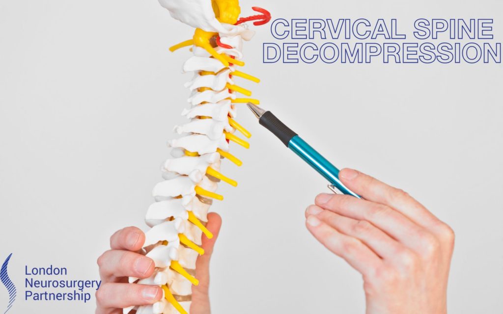
Mr Richard Gullan one of the London Neurosurgery Partnership cervical spine specialists discusses cervical spine decompression surgery…
The cervical area of the spine refers to the neck region of the spine. This includes the very topmost bones of the spine – C1 and C2 (which the head sits on, down to C7 – the bottommost bone of the cervical region. There are 6 cervical discs between the vertebrae (cervical bones) and 8 pairs of cervical nerves exiting the spinal cord to supply the arms and shoulders. Cervical spine decompression surgery is in the cervical region of the spine.
Decompression surgery refers to the opening up of the spinal canal or the foramen (through which the nerve roots exit the spinal cord). Decompression surgery is usually performed if there is a narrowing of the spinal canal or foramen causing compression of the nerves. This can lead to chronic pain, numbness, tingling and muscle weakness (see blog on cervical spine stenosis). The aim of decompression surgery is to relieve the pressure on the affected nerves to restore or preserve function. Surgery is usually only recommended if your symptoms have not improved with physical therapy or medications or if symptoms are extremely severe. Cervical spine decompression surgery is decompression surgery in the cervical region of the spine.
Cervical spine decompression surgery is performed via an incision in the back (posterior) muscles of the neck. The lamina (which is the back portion of bone of the spinal canal, it essentially forms a roof over the spinal cord) is removed or partially removed during a decompression. By removing the lamina and any thickened spinal ligament the rear wall of the box encasing the cervical spinal cord and nerves is taken off to give more room for the nerves. This also allows for the removal of any bony spurs (called osteophytes) which may be pinching the nerves. The surgeon may address one vertebra (single-level) or more (multi-level) during decompression surgery depending on the symptoms the patient is experiencing .
Decompression surgery is the umbrella term for several different types of surgery which relieves pressure on the nerves, it just depends on the level of compression and therefore how much bone needs to be removed:
- Laminectomy – the surgeon will remove the entirety of the lamina plus some of the facet joint and any thickened ligament.
- Laminotomy – this operations sees a smaller portion of the lamina and ligaments removed, this is often just on one side of the neck. By removing less bone there is less chance of any neck instability.
- Foraminotomy – this is the removal of bone from just around the neural foramen (the channel where the nerves exit the spinal to supply the upper limbs). Surgeons may adopt this method when the cervical disc is the cause of the nerve compression. This is usually performed on one side and involves a small incision and less muscle disruption that a laminectomy.
- Laminaplasty – refers to the expansion of the cervical spinal canal by the cutting the lamina on one side and hinging it open, before fixing it open with small plate.
- Discectomy – this is the removal of a piece of disc which is causing compression of a nerve root. This could be a disc bulge/herniation or degenerative disc.
- Microdiscectomy – the aim of the surgery is to remove parts of the herniated disc from the neck and alleviate pain and other symptoms using a keyhole, minimally invasive approach.
Sometimes, a spinal fusion may be done at the same time as cervical spine decompression surgery to help stabilise the area of the neck where the lamina was removed. Fusion uses a combination of bone graft, screws, and rods to connect two separate vertebrae together into one new piece of bone. Fusing the joint prevents the spinal stenosis from recurring and can help eliminate pain from an unstable spine.
This article is intended to inform and give insight but not treat, diagnose or replace the advice of a doctor. Always seek medical advice with any questions regarding a medical condition.

0 Comments