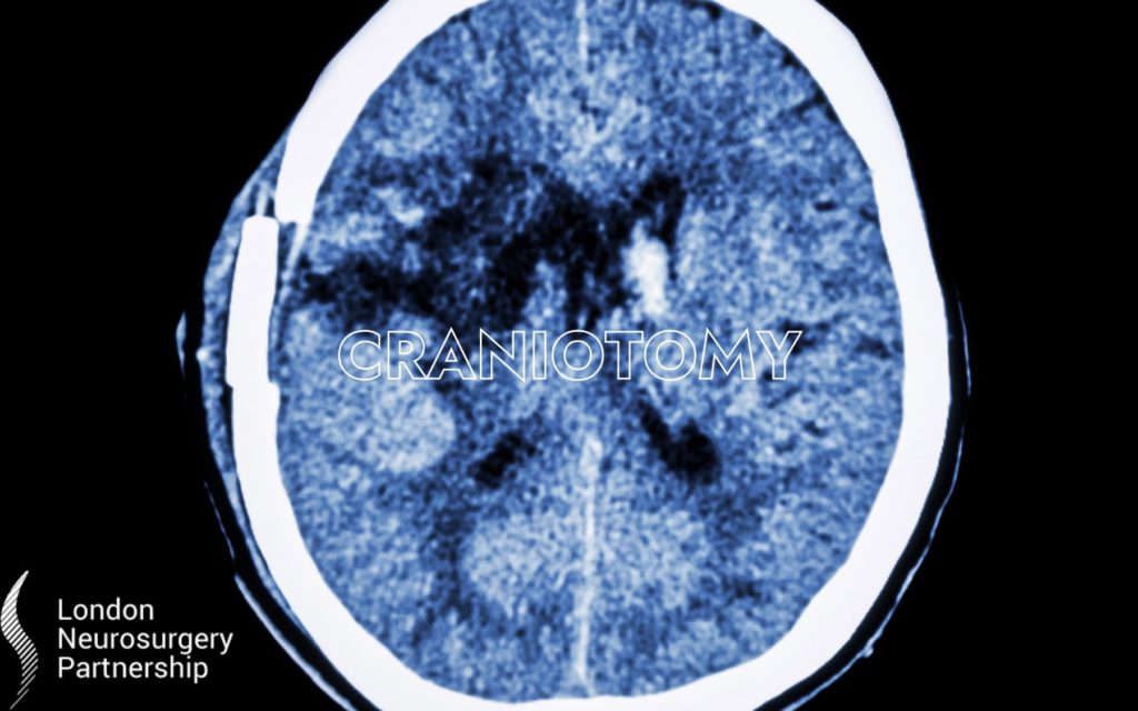
What is a craniotomy?
A craniotomy is the most common type of operation to remove a brain tumour. To access the tumour, the surgeon will cut out an area of bone from the skull which is referred to as a ‘flap’. The surgeon will use scans done prior to the operation to accurately locate and remove as much of the tumour as possible, without taking any healthy tissue. The ‘flap’ is then replaced and secured with small permanent metal brackets. The scalp is stitched back into place, and in most cases, this will be covered by the patient’s hair.
Depending on the location of the brain tumour, the surgeon might suggest an ‘awake craniotomy’. If the tumour is close to part of the brain that controls an important function eg, speech, the surgeon may want to ask the patient to do tasks eg., speak, whilst checking the function of the brain. There may be the option to have general anaesthetic for part of the operation which will be discussed with your surgeon. Either way, a local anaesthetic will be used for any parts of the surgery when the patient is awake so they will not feel any pain.
An interesting fact about the brain is that the brain itself does not feel pain, therefore although an awake craniotomy may seem daunting, the team around you will ensure you are not in any pain.
Craniotomies are also used to treat subdural haematomas, extradural haematomas, meningiomas, gliomas and glioblastomas. Craniotomies can also, in certain circumstances be performed as awake brain surgery.
Minimally invasive technique: Neuroendoscopy
Neuroendoscopy is a minimally invasive technique, sometimes referred to as keyhole, which requires a much smaller opening in the skull. An endoscope which is made up of a long tube, camera and small surgical instruments is passed through this small opening. The camera projects the image inside the brain on to a TV screen, the surgeon will use this as a guide to remove the tumour. The benefit of this method is that it is less invasive and allows for a faster recovery. This type of surgery is particularly useful for removing ependymoma’s in the ventricles (the fluid filled spaces of the brain).
This article is intended to inform and give insight but not treat, diagnose or replace the advice of a doctor. Always seek medical advice with any questions regarding a medical condition.
0 Comments