Mr Francesco Vergani, our resident TMS specialist, in this week’s blog discusses all about TMS or transcranial magnetic stimulation.
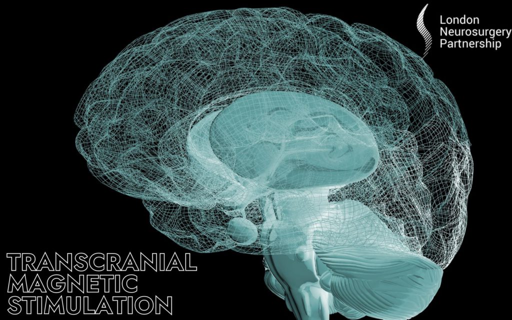
Transcranial magnetic stimulation (TMS) is a non-invasive procedure that uses magnetic fields to stimulate nerve cells in the brain. This new technique has advanced the understanding of brain physiology, and has diagnostic value as well as therapeutic potential for several neuropsychiatric disorders.
The term TMS can be confusing as the machinery used doesn’t actually contain a magnet. It is in fact a coil of wire which sends out a magnetic field. The magnetic field then transmits a small electrical current through the brain. A TMS electric field affects only a very small part of the total brain area. The TMS electric current can reach all the way down through your cortex.
The stimulation involves restricted cortical and subcortical regions, and, when used in combination with a visually guided technique, results in improved accuracy to target specific areas, which can help the surgeon to navigate the brain tissue through defined “safe tracts”. TMS can be used to obtain functional maps of the eloquent areas of the brain, the motor and visual cortex, which can be used for surgical planning.
One of the great things about TMS is that its effects are only on the brain. It is completely non-invasive and considered safe. There is no need for a needle or swallowing pills; magnetic fields simply pass through brain tissue, harmlessly.
So how does TMS actually work?
The brain cells are called neurons, they transmit electrical impulses to send messages through the brain – so communication in the brain is based on electricity which ultimately, is the same electricity that TMS generates. TMS works because each magnetic pulse made by the TMS creates a local electric field in the brain which is just strong enough to activate the neurons needed. As these neurons are now forced into activation, in turn they then activate other, neighbouring neurons and strengthen connections to other parts of the brain. It stimulates the brain to work!
The equipment we use at London Neurosurgery Partnership is called Nexstim SmartFocus® nTMS and it allows us to plan and execute our most delicate procedures, from tumours to AVM lesions in the most eloquent areas. We use TMS after a patient is diagnosed with a brain tumour, or another disorder and when the lesion is thought to be close to vital areas of the brain, such as those responsible for limb movement or speech production.
It is important that the patient is diagnosed first, as we are able to understand more about the type and grade of the tumour or brain lesion, then most crucial information needed for a treatment decision is its location in relation to the brain’s vital functions and their sub-cortical connections. We also need to understand the exact locations of vital brain functions so we can determine operability and plan the safest approach for the best outcome possible whilst minimizing any risk of post-operative deficits.
Working with Nexstim SmartFocus® nTMS map allows an accurate and reliable picture which allows us and the patient discuss the best treatment options for their case as well as discussing the risks of a gross total resection vs debulking.
SmartFocus® nTMS motor mapping is suitable for children, as well as for people with special needs, and can be successful even when the patient has hemiplegia.
This article is intended to inform and give insight but not treat, diagnose or replace the advice of a doctor. Always seek medical advice with any questions regarding a medical condition.
0 Comments