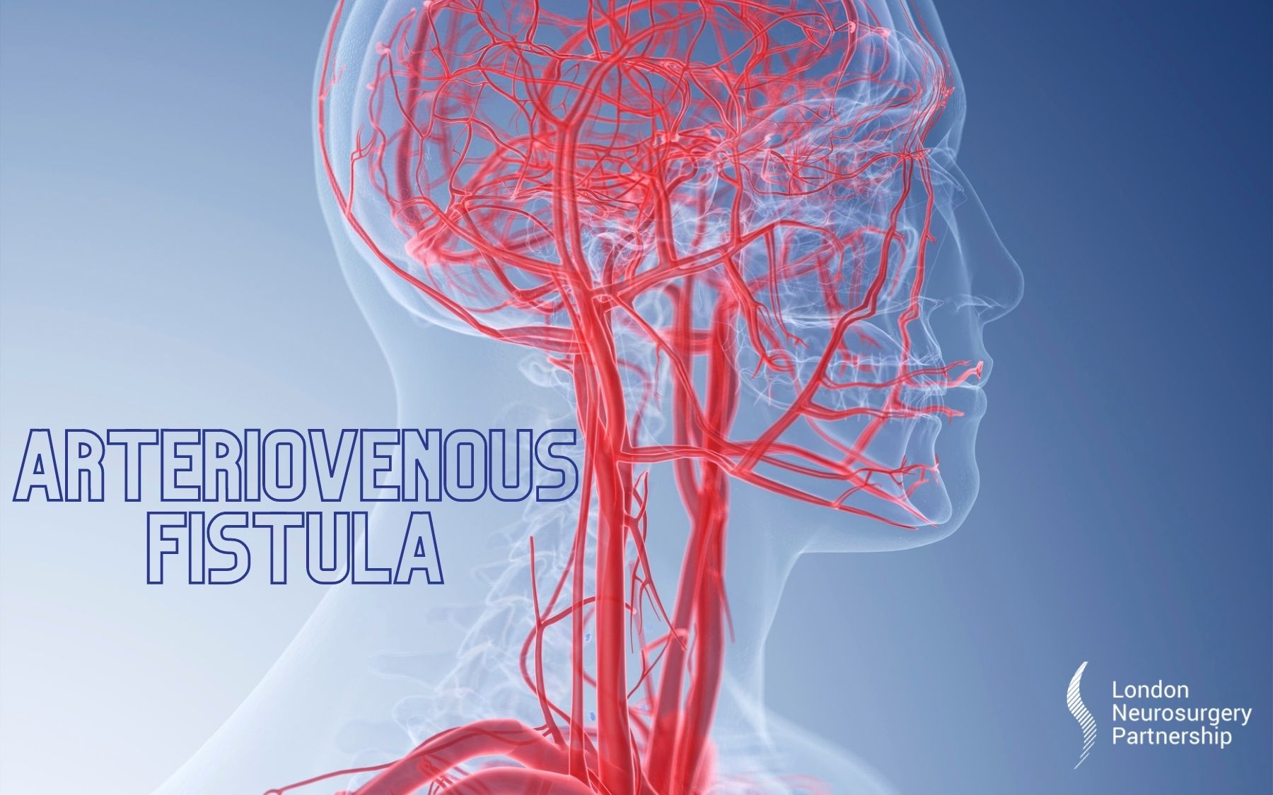

An arteriovenous fistula is a neuro condition that is caused by an abnormal connection between an artery and a vein. Normally, blood flows from your arteries to your capillaries, and then on to your veins. Nutrients and oxygen in your blood travel from your capillaries to tissues in your body. With an arteriovenous fistula, blood flows directly from an artery into a vein, bypassing some capillaries. When this happens, tissues below the bypassed capillaries receive less blood. Arteriovenous fistulae differ from arteriovenous malformations (AVMs) in that AVMs are found within the tissue of the brain or spinal cord, but AV fistulae are found in the coverings of the brain or spinal cord, such as the dura mater or arachnoid. The most serious problem associated with AV fistulae is that they transfer high-pressure arterial blood into the veins that drain blood from the brain or spinal cord. This results in an increase in the pressure of the venous system around the brain or spinal cord.
An arteriovenous fistula can occur both in and around the brain, as well as the spine. An arteriovenous fistula can be present at birth (congenital), develop after birth or develop due to an injury (acquired)
Here is a list of some of the most common types of arteriovenous fistula:
Acquired arteriovenous fistula
These are not present at birth. They usually happen when a sharp object goes through your body tissue, such as during a gunshot or stabbing injury.
Dural arteriovenous fistula
This type of arteriovenous fistula occurs within the dura. This is the layer that covers the brain and spinal cord. In some cases, this condition will be picked up at birth, whereas others may not be noticed until later in childhood. A large dural AV fistula can cause cardiac failure at birth. Smaller ones can cause increase in the pressure in the veins inside the head, resulting in hydrocephalus (a build-up of fluid in the brain) or an enlarged cerebral ventricle (spaces within the brain that hold fluid). A dural AV fistula can also cause a pulsing noise or less often, bleeding or damage to the brain tissue.
Pial or cerebral arteriovenous fistula
This occurs in the brain. A large one can cause heart failure at birth or before birth. Smaller ones can damage the brain around the fistula because it diverts blood flow away from the brain tissue and into the draining vein. If this condition is detected it is treated straight away to minimise the amount brain damage that occurs.
Spinal arteriovenous fistula
This occurs in or around the spinal cord, within the spine or in the muscles near to the spine. These fistulas can compress the spinal cord, leading to symptoms such as numbness or weakness.
Vein of Galen arteriovenous fistula
These usually appear in infancy or early childhood. They drain into the vein of Galen, which is part of the deep venous drainage system of the brain. These fistulas can cause cardiac failure, hydrocephalus, or damage to the developing brain.
Early detection of an arteriovenous fistula may make your condition easier to treat. It also may reduce your risk of developing complications, including blood clots or heart failure.
Symptoms
There are two major types of AVFs we see in neurosurgery: dural AVFs and carotid-cavernous fistulas (CCFs). These are both acquired lesions, which means that patients are not born with them, but instead develop them later in life. They can be a result of infection or traumatic injuries, but most develop without any specific precipitating event. Patients with dural AVFs typically present with a rumbling noise in one ear that follows the heartbeat, which is called a bruit. Patients with CCFs typically present with swelling and redness of one or both eyes in addition to a bruit.
Diagnosis
Currently, we aim whenever possible to close the AV fistula before the increased pressure in the venous system causes irreversible damage to the brain or spinal cord. We usually diagnose AV fistulae with an angiogram. An angiogram is a test in which a neuroradiologist injects dye into the blood vessels in the brain to obtains images of the network of blood vessels.
Treatment
- Minimally invasive endovascular embolization — usually this sufficient to cure the AV fistula. During this procedure, a catheter is fed through the groin up into the arteries in the brain that supply the AV fistula and inject liquid embolic agents into these arteries. This injection shuts off that artery and reduces the flow of blood to the fistula.
- Microsurgical resection — this is used solely for AV fistulae which cannot be treated with endovascular embolization. During microsurgical resection, we perform a craniotomy and using the microscope, isolate and remove the AV fistula from the tissues around the brain or spinal cord.
If you would like to speak to Mr Tolias, our AV fistula and neurovascular specialist, about diagnosis or treatment for an arteriovenous fistula, please call us on 0207 034 8709 or email us at info@londonneurosurgerypartnership.co.uk

0 Comments