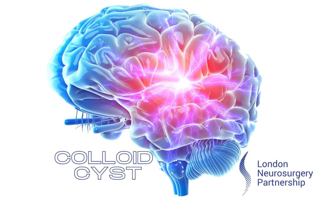
A colloid cyst is a benign brain tumour, which means it’s non-cancerous. Mr Christopher Chandler explains to us what actually is a colloid cyst. They do not spread but will slowly grow in size. Colloid cysts are small fluid-filled sacs located in or around the lateral and third ventricle of the brain. Because of its location in the ventricles, a colloid cyst can sometimes cause a blockage of cerebral spinal fluid (CSF). CSF is located in the ventricles where is protects and cushions the brain and spinal cord, if the flow of CSF is interrupted it can cause a person to develop hydrocephalus (excess CSF in the brain) as the colloid cyst is disrupting the body’s natural circulation of fluid.
There is no defining cause why a colloid cyst develops but it is thought to be present from foetal development. They do tend to grow as a person grows in to an adult as it is rare to find a colloid cyst in a child.
Symptoms
Colloid cysts are usually diagnosed through incidental findings. This means that it was found almost by accident, maybe when a patient was having a scan for a different reason like headaches. This is because they do not often present with symptoms as they are slow growing, the brain has time to get used to it being there.
A colloid cyst will start to cause symptoms when it begins to block the CSF flow the circulate the brain and spine. This will lead to hydrocephalus. Symptoms of this will include headaches and sometimes vomiting, visual disturbances, memory problems and in extreme cases loss of consciousness and coma.
Diagnosis
If your doctor suspects your symptoms are being causes by something in the brain then they will investigate by asking the patient to have an MRI scan. This scan is will used specific sequences to capture images of the brain and spinal cord. They are able to identify colloid cysts. This is the quickest way to diagnose a colloid cyst. A doctor is able to see the imaging immediately after the scan is performed.
Treatment
Treatment for a colloid cyst will differ from person to person as it all depends on the age of the patient, location, size and severity of the cyst and if it is causing CSF blockage.
If a colloid cyst is found to be causing no symptoms or hydrocephalus, is small and is not affecting the patient’s life then a doctor may recommend a watch and wait technique. This means there is not a good enough reason to operate on the patient as its not affecting the brain. The doctor will be able to monitor the cyst with MRI scanning annually. If the cyst is growing in size and the patient starts to present with symptoms then surgical removal may be an option. If not, then the monitoring will continue.
If the cyst is interrupting the natural CSF flow of the brain and causing hydrocephalus then surgical intervention will be necessary. This will be in the form of an endoscopic craniotomy – a minimally invasive technique. The cyst will be carefully drained and then resected from the brain. If the hydrocephalus does not resolve then a shunt device will need to be inserted to drain the excess fluid from the brain.
This article is intended to inform and give insight but not treat, diagnose or replace the advice of a doctor. Always seek medical advice with any questions regarding a medical condition.
Back to brain conditions.


0 Comments