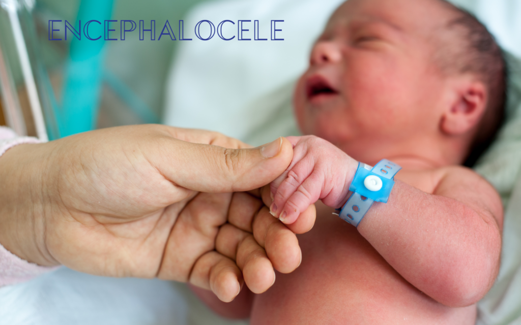
Encephalocele is a rare congenital birth defect, where the skull does not form properly which allows brain tissue, cerebrospinal fluid and blood vessels to protrude outside the brain. Occurring during foetal development, encephalocele has a protruding external sac-like appearance that is covered either in skin or thin membrane. The encephalocele can open anywhere along the centre of the skull, which means the sac can protrude from the back of the head to the forehead and nose, if it forms on the face it is called a midfacial cleft.
According to Shine Charity – Occipital encephaloceles are more common in Europe and North America, whereas anterior encephaloceles are more common in Southeast Asia, Africa, Malaysia and Russia. They occur in 1 in every 5,000 births.
During early development, the brain and spinal cord form in the shape of a tube that is open at either end, this is called a neural tube. As you may know, the first few weeks of pregnancy are extremely important to the baby’s initial development and as the neural tube continues to grow, the brain and spinal cord begin to form.
Why does encephaloceles occur?
It can be possible for the neural tube to not close and develop properly which can cause neural tube defects. We do not know the exact reason as to why encephaloceles happen and in most cases, it happens sporadically, but it is possible to have an increased risk if there is a family history in neural tube defects.
It is known and advised that folic acid is to be taken throughout pregnancy as it can help reduce the risk of encephaloceles and other deformities occurring.
In most cases, encephalocele is diagnosed at birth but in some cases, it can be seen on prenatal ultrasound and therefore a caesarean section will be planned as this will be the safer route as opposed to giving birth vaginally.
Diagnosing encephalocele
Further imaging scans may be necessary to diagnose encephalocele such as MRI. MRI scanning will take a more in-depth look at the skull and will be able to determine whether the fluid-filled sac contains and tissue or meninges.
It is possible to treat encephalocele but it does depend on the size, the contents of the sac and how it will affect the baby. During foetal development, if the encephalocele grows too large, that baby may not survive the pregnancy. If it continues to stay small and brain tissue does not protrude into the sac, then it is possible for a good neurosurgical outcome.
When blood vessels and brain tissue protrude into the sac, surgical intervention can be more difficult and an MDT (multi-disciplinary meeting) may need to take place to decide whether surgery is the best option.
Treatment for encephalocele
Surgery is required to treat encephalocele and it will be performed under general anaesthetic. Surgery timing can vary from after birth to a couple of months after. This is because if the sac is covered with a thin membrane, then surgery after birth will be the best option to prevent infection or any damage. If there is skin covering the sac then surgery can be delayed to allow the baby to grow and develop.
There may be a build-up of cerebrospinal fluid (hydrocephalus) in which case a shunt will be inserted to drain the excess fluid into the abdomen. When operating on the encephalocele, any brain tissue or meninges will need to be repositioned back into the skull, this will be done by cutting through the dura and removing a small part of the skull. The skull and the dura will then be repaired or replaced and then can be closed. incision closed.
Neurosurgical observations will take place to check that the shunt is draining the excess CSF fluid currently and this will be closely monitored alongside the baby’s recovery.
If you would like further information, we have paediatric neurosurgeons Mr Sanj Bassi and Mr Christopher Chandler who are committed to treating babies and children born with congenital defects.
This article is intended to inform and give insight but not treat, diagnose or replace the advice of a doctor. Always seek medical advice with any questions regarding a medical condition.


0 Comments