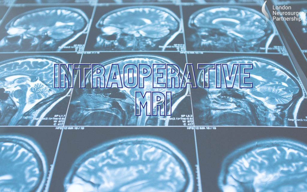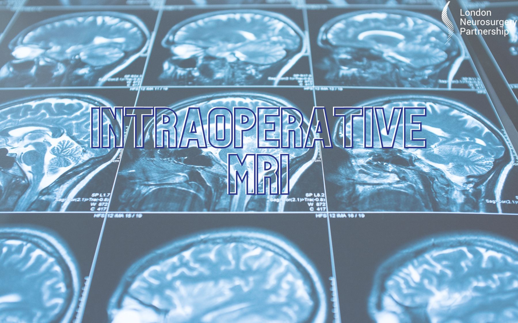
Intraoperative magnetic resonance imaging, intraoperative MRI or iMRI is a technology that uses an MRI scanner to create images of the brain during surgery. MRI scans work in a completely different way to CT scans and X-ray’s; they use both radio waves and strong magnets. Once the MRI scan has been completed, software is used to create a 3D image that shows the different tissue types -this is a great scan for imaging soft tissue such as the brain, blood vessels, spinal discs and spinal cord. An MRI scan is commonly used to diagnose problems with the tissue, such as discs, muscle problems, blood flow problems and tumours. iMRI brings this technology into the operating theatre. Surgeons use iMRI to assist in procedures that treat a variety of brain conditions including brain tumours, epilepsy, movement disorders and vascular conditions such as cavernomas and tangled blood vessels (arteriovenous malformation).
The use of iMRI during surgery allows surgeons to monitor and image the structure and functional connections of the brain during the operation; they can check they have achieved maximal safe removal of lesions; check for bleeding, clots and other complications; prevent damage to surrounding tissue; and protect brain function.
What are the advantages of iMRI?
iMRI allows the surgeons to ensure that they have completely removed the lesion which means that they don’t have to go back for a second surgery a few days later. For example, complete tumour removal is the best predictor of increased life expectancy.
iMRI identifies any intra-operative (during the surgery) complications before surgery is finished (e.g. a hidden blood clot) so allowing these to be dealt with before they cause harm and preventing the need for a second operation.
The brain often moves during surgery, which makes pre-surgical imaging not exactly precise. Imaging with iMRI during the operation helps to provide surgeons with the most accurate information as the images are taken in real-time so the surgeons can locate abnormalities if the brain has shifted at all during the procedure.
It can be difficult to clearly identify the very edge of a brain lesion, especially some types of tumour, and separate normal, heathy brain tissue from abnormal tissue. Using iMRI to generate images during surgery helps confirm successful removal of the entire brain lesion.
Professor Ashkan: “The neurosurgical multidisciplinary team at the LNP is delighted to announce access to another cutting edge surgical technology. Intra-operative MRI (iMRI) will further aid efficacy of brain surgery by helping to maximise to extent of surgical resection and also the safety of surgery. Combining iMRI with advanced techniques we already use, such as brain mapping, awake surgery and intra operative fluorescence, means that our patients can be reassured that every advanced technique available anywhere in the world is now available to them.”


0 Comments