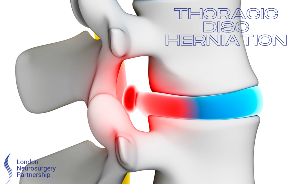
A thoracic disc herniation can be a painful condition where the fibrous outer part of the disc tears and the inner gelatinous material leaks out from inside the disc. A thoracic herniated disc can cause upper back and neck pain that can radiate to other parts of your body. When the herniated disc ruptures it can compress a nearby nerve resulting in a pinched nerve, inflaming and irritating the nerve causing additional pain such as tingling, numbness and weakness.
Our spinal columns are made up of 33 bones called vertebra. Intervertebral discs are strong connective tissues that separate each vertebra. Ligaments and facet joints help connect the individual vertebra to maintain movement, curvature and the spines normal alignment.
The word ‘hernia’ means when an internal part of the body pushes out through a weakness in the surrounding tissue or muscle, causing a hernia. The word ‘disc’ means the spongy cushions that sperate the bones from the spine, hence the name of a thoracic disc herniation.
Thoracic disc herniation can be caused by trauma from a possible sports injury or accident. It is the sudden and strong force that can lead to a herniated disc. Other causes are possible from mild trauma such as a work accident, reaching up high whilst twisting or even a fall and usually the symptoms will worsen over time or from a degenerative disc if the patient is older in age.
It is possible for a herniated disc not to present with any signs or symptoms at all. Most symptoms are a result of increased pressure and irritation on the nerves. Some of the symptoms associated with thoracic disc herniation are:
- Muscle weakness in one or both legs
- Numbness in one or both legs
- Tingling in one or both legs
- Radiating pain in one or both legs
- Spasticity in the legs
In some cases, there is also the possibility of total paralysis in the legs. The above symptoms will differ from patient to patient and are suggestive to which nerve has been affected and how much pressure is on the spinal cord.
Diagnosing a thoracic disc herniation begins with a consultation with your doctor. You will give information about your medical history, symptoms and what you think is the possible cause for your pain. You will then have a physical examination that will test movements such as your reflexes and balance.
For a definitive diagnosis, an MRI or X-ray will be conducted. An MRI scan is a definitive way of diagnosing conditions, It is able to pick up detailed images of your spine in specific sequences, helping the consultant find the source of your pain,
Treatment
Treatment for a thoracic disc herniation ultimately depends on its seriousness and associated symptoms. It is possible for symptoms to get better over time and the thoracic herniated disc to completely resolve by itself. In this case just resting and observing may be the best option, making sure there is no progression in symptoms. If symptoms do happen to get worse then there are treatments designed to help relieve and take care of the condition.
Medication is another surgical free option offered to patients when suffering from a thoracic disc herniation. Pain-relief such as ibuprofen, Tylenol and anti-inflammatory medications are designed to reduce pain. It is important to always take your doctor’s advice when taking medication and to never take more than recommended. If the pain is not getting any better than the best option is always to speak with a doctor first.
Treatments such as an Epidural Steroid Injection (EPI) is a fast and almost immediate way to reduce the pain of a thoracic disc herniation for a certain amount of time, it is usually suggested if all other conservative routes have not worked and surgical intervention is fast becoming an option.
Lastly, surgery can be an option for those thoracic disc herniations that are becoming considerably worse. A laminotomy or discectomy is a traditional way of treating a herniated disc.
Performed under general anaesthetic the aim of the surgery is to remove any ruptured disc material and allow space for the compressed nerves. During the procedure, an incision is made through the skin, and muscles are moved to the side to expose the vertebrae. Using X-ray assistance, an opening is made between the two correct vertebrae to locate the ruptured disc. Now the spinal canal is in view, a laminotomy may need to take place, this means some of the bone from the lamina may need to be removed to allow space for the procedure to take place. The surgeon will be able to locate the disc material and remove it, this will remove any pressure from the nerves. The surgeon may then remove any nucleus pulposus material to prevent any further hernias in the future.
A microdiscectomy is another procedure to correct a thoracic herniated disc, this is more of a minimally invasive procedure using special tools through a much smaller incision. Some advantages of this procedure are less risk of infection and quicker recovery times.
If you would like further information on the surgical procedures we offer for thoracic herniated disc, please give our office a call
This article is intended to inform and give insight but not treat, diagnose or replace the advice of a doctor. Always seek medical advice with any questions regarding a medical condition.

0 Comments